

How To Draw Diagram Of Gram Positive Cell Wall
In bacteriology, gram-positive bacteria are leaner that give a positive effect in the Gram stain test, which is traditionally used to quickly allocate bacteria into two broad categories co-ordinate to their blazon of cell wall.
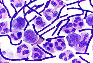
Gram-positive bacteria take up the crystal violet stain used in the test, and and then appear to exist imperial-coloured when seen through an optical microscope. This is considering the thick peptidoglycan layer in the bacterial cell wall retains the stain after it is done away from the residual of the sample, in the decolorization stage of the test.
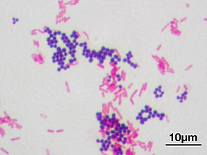
Conversely, gram-negative bacteria cannot retain the violet stain after the decolorization step; alcohol used in this stage degrades the outer membrane of gram-negative cells, making the cell wall more porous and incapable of retaining the crystal violet stain. Their peptidoglycan layer is much thinner and sandwiched between an inner prison cell membrane and a bacterial outer membrane, causing them to take up the counterstain (safranin or fuchsine) and appear ruby-red or pink.
Despite their thicker peptidoglycan layer, gram-positive bacteria are more than receptive to certain cell wall targeting antibiotics than gram-negative bacteria, due to the absenteeism of the outer membrane.[i]
Characteristics [edit]
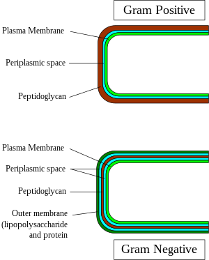
Gram-positive and gram-negative jail cell wall construction

Structure of gram-positive cell wall
In general, the following characteristics are nowadays in gram-positive bacteria:[2]
- Cytoplasmic lipid membrane
- Thick peptidoglycan layer
- Teichoic acids and lipoids are nowadays, forming lipoteichoic acids, which serve as chelating agents, and also for certain types of adherence.
- Peptidoglycan chains are cross-linked to form rigid cell walls by a bacterial enzyme DD-transpeptidase.
- A much smaller book of periplasm than that in gram-negative bacteria.
Just some species have a capsule, usually consisting of polysaccharides. Also, merely some species are flagellates, and when they practise have flagella, have simply two basal body rings to support them, whereas gram-negative have four. Both gram-positive and gram-negative bacteria commonly have a surface layer called an S-layer. In gram-positive bacteria, the Due south-layer is attached to the peptidoglycan layer. Gram-negative bacteria's Southward-layer is attached directly to the outer membrane. Specific to gram-positive bacteria is the presence of teichoic acids in the cell wall. Some of these are lipoteichoic acids, which have a lipid component in the cell membrane that tin can assist in anchoring the peptidoglycan.
Classification [edit]
Along with cell shape, Gram staining is a rapid method used to differentiate bacterial species. Such staining, together with growth requirement and antibody susceptibility testing, and other macroscopic and physiologic tests, forms the total basis for nomenclature and subdivision of the leaner (eastward.1000., meet figure and pre-1990 versions of Bergey's Manual).
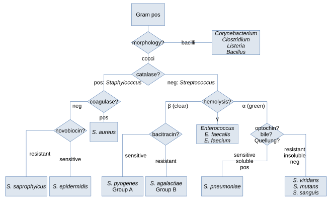
Species identification hierarchy in clinical settings
Historically, the kingdom Monera was divided into 4 divisions based primarily on Gram staining: Firmicutes (positive in staining), Gracilicutes (negative in staining), Mollicutes (neutral in staining) and Mendocutes (variable in staining).[3] Based on 16S ribosomal RNA phylogenetic studies of the late microbiologist Carl Woese and collaborators and colleagues at the University of Illinois, the monophyly of the gram-positive bacteria was challenged,[four] with major implications for the therapeutic and full general report of these organisms. Based on molecular studies of the 16S sequences, Woese recognised twelve bacterial phyla. Two of these were gram-positive and were divided on the proportion of the guanine and cytosine content in their DNA. The high G + C phylum was fabricated up of the Actinobacteria and the low G + C phylum contained the Firmicutes.[4] The Actinobacteria include the Corynebacterium, Mycobacterium, Nocardia and Streptomyces genera. The (low G + C) Firmicutes, take a 45–60% GC content, but this is lower than that of the Actinobacteria.[2]
Importance of the outer cell membrane in bacterial nomenclature [edit]

Although bacteria are traditionally divided into 2 chief groups, gram-positive and gram-negative, based on their Gram stain retention holding, this classification system is ambiguous equally it refers to iii distinct aspects (staining consequence, envelope organization, taxonomic group), which do non necessarily coalesce for some bacterial species.[5] [half-dozen] [7] [8] The gram-positive and gram-negative staining response is also not a reliable feature as these two kinds of bacteria do not course phylogenetic coherent groups.[5] Withal, although Gram staining response is an empirical criterion, its basis lies in the marked differences in the ultrastructure and chemical composition of the bacterial cell wall, marked by the absenteeism or presence of an outer lipid membrane.[5] [ix]
All gram-positive bacteria are bounded past a single-unit lipid membrane, and, in general, they contain a thick layer (twenty–80 nm) of peptidoglycan responsible for retaining the Gram stain. A number of other bacteria—that are bounded by a single membrane, merely stain gram-negative due to either lack of the peptidoglycan layer, as in the mycoplasmas, or their inability to retain the Gram stain because of their jail cell wall composition—also show close relationship to the Gram-positive bacteria. For the bacterial cells divisional by a single cell membrane, the term monoderm bacteria has been proposed.[5] [9]
In contrast to gram-positive bacteria, all typical gram-negative leaner are divisional by a cytoplasmic membrane and an outer cell membrane; they contain only a sparse layer of peptidoglycan (2–iii nm) between these membranes. The presence of inner and outer cell membranes defines a new compartment in these cells: the periplasmic infinite or the periplasmic compartment. These bacteria have been designated as diderm bacteria.[v] [9] The distinction between the monoderm and diderm bacteria is supported by conserved signature indels in a number of important proteins (viz. DnaK, GroEL).[five] [half-dozen] [ix] [10] Of these 2 structurally singled-out groups of bacteria, monoderms are indicated to be ancestral. Based upon a number of observations including that the gram-positive bacteria are the major producers of antibiotics and that, in general, gram-negative leaner are resistant to them, it has been proposed that the outer prison cell membrane in gram-negative leaner (diderms) has evolved every bit a protective machinery confronting antibiotic selection pressure.[5] [six] [9] [10] Some bacteria, such as Deinococcus, which stain gram-positive due to the presence of a thick peptidoglycan layer and also possess an outer cell membrane are suggested as intermediates in the transition betwixt monoderm (gram-positive) and diderm (gram-negative) bacteria.[5] [ten] The diderm leaner can also be further differentiated between simple diderms lacking lipopolysaccharide, the archetypical diderm bacteria where the outer cell membrane contains lipopolysaccharide, and the diderm leaner where outer cell membrane is made up of mycolic acid.[vii] [ten] [11]
Exceptions [edit]
In general, gram-positive bacteria are monoderms and have a single lipid bilayer whereas gram-negative bacteria are diderms and have two bilayers. Some taxa lack peptidoglycan (such equally the course Mollicutes, some members of the Rickettsiales, and the insect-endosymbionts of the Enterobacteriales) and are gram-variable. This, however, does not e'er hold true. The Deinococcus-Thermus bacteria have gram-positive stains, although they are structurally similar to gram-negative leaner with two layers. The Chloroflexi take a single layer, yet (with some exceptions[12]) stain negative.[thirteen] Two related phyla to the Chloroflexi, the TM7 clade and the Ktedonobacteria, are as well monoderms.[xiv] [fifteen]
Some Firmicute species are not gram-positive. These vest to the class Mollicutes (alternatively considered a class of the phylum Tenericutes), which lack peptidoglycan (gram-indeterminate), and the class Negativicutes, which includes Selenomonas and stain gram-negative.[eleven] Additionally, a number of bacterial taxa (viz. Negativicutes, Fusobacteria, Synergistetes, and Elusimicrobia) that are either part of the phylum Firmicutes or co-operative in its proximity are constitute to possess a diderm cell structure.[viii] [10] [xi] Nonetheless, a conserved signature indel (CSI) in the HSP60 (GroEL) protein distinguishes all traditional phyla of gram-negative bacteria (e.thousand., Proteobacteria, Aquificae, Chlamydiae, Bacteroidetes, Chlorobi, Cyanobacteria, Fibrobacteres, Verrucomicrobia, Planctomycetes, Spirochetes, Acidobacteria, etc.) from these other atypical diderm bacteria, as well as other phyla of monoderm bacteria (e.g., Actinobacteria, Firmicutes, Thermotogae, Chloroflexi, etc.).[10] The presence of this CSI in all sequenced species of conventional LPS (lipopolysaccharide)-containing gram-negative bacterial phyla provides testify that these phyla of bacteria course a monophyletic clade and that no loss of the outer membrane from whatsoever species from this group has occurred.[10]
Pathogenesis [edit]

Colonies of a gram-positive pathogen of the oral crenel, Actinomyces sp.
In the classical sense, half dozen gram-positive genera are typically pathogenic in humans. Two of these, Streptococcus and Staphylococcus, are cocci (sphere-shaped). The remaining organisms are bacilli (rod-shaped) and can be subdivided based on their ability to form spores. The non-spore formers are Corynebacterium and Listeria (a coccobacillus), whereas Bacillus and Clostridium produce spores.[xvi] The spore-forming leaner can again exist divided based on their respiration: Bacillus is a facultative anaerobe, while Clostridium is an obligate anaerobe.[17] Likewise, Rathybacter, Leifsonia, and Clavibacter are three gram-positive genera that cause found disease. Gram-positive bacteria are capable of causing serious and sometimes fatal infections in newborn infants.[18] Novel species of clinically relevant gram-positive bacteria as well include Catabacter hongkongensis, which is an emerging pathogen belonging to Firmicutes.[19]
Bacterial transformation [edit]
Transformation is one of iii processes for horizontal factor transfer, in which exogenous genetic textile passes from a donor bacterium to a recipient bacterium, the other ii processes being conjugation (transfer of genetic cloth between 2 bacterial cells in directly contact) and transduction (injection of donor bacterial DNA by a bacteriophage virus into a recipient host bacterium).[20] In transformation, the genetic material passes through the intervening medium, and uptake is completely dependent on the recipient bacterium.[twenty]
Every bit of 2014 about lxxx species of leaner were known to be capable of transformation, near evenly divided between gram-positive and gram-negative bacteria; the number might exist an overestimate since several of the reports are supported by single papers.[20] Transformation among gram-positive bacteria has been studied in medically important species such as Streptococcus pneumoniae, Streptococcus mutans, Staphylococcus aureus and Streptococcus sanguinis and in gram-positive soil bacterium Bacillus subtilis, Bacillus cereus.[21]
Orthographic note [edit]
The adjectives Gram-positive and Gram-negative derive from the surname of Hans Christian Gram; every bit eponymous adjectives, their initial letter can be either capital G or lower-instance g, depending on which manner guide (e.g., that of the CDC), if whatsoever, governs the certificate existence written.[22] This is further explained at Gram staining § Orthographic note.
References [edit]
- ^ Basic Biology (18 March 2016). "Bacteria".
- ^ a b Madigan, Michael T.; Martinko, John M. (2006). Brock Biology of Microorganisms (11th ed.). Pearson Prentice Hall. ISBN978-0131443297.
- ^ Gibbons, North. East.; Murray, R. G. E. (1978). "Proposals Concerning the Higher Taxa of Bacteria". International Journal of Systematic and Evolutionary Microbiology. 28 (1): 1–6. doi:10.1099/00207713-28-1-1.
- ^ a b Woese, C. R. (1987). "Bacterial evolution". Microbiological Reviews. 51 (2): 221–271. doi:ten.1128/MMBR.51.2.221-271.1987. PMC373105. PMID 2439888.
- ^ a b c d eastward f 1000 h Gupta, R. S. (1998). "Protein phylogenies and signature sequences: A reappraisal of evolutionary relationships among archaebacteria, eubacteria and eukaryotes". Microbiology and Molecular Biology Reviews. 62 (4): 1435–1491. doi:x.1128/MMBR.62.4.1435-1491.1998. PMC98952. PMID 9841678.
- ^ a b c Gupta, R. S. (2000). "The natural evolutionary relationships amid prokaryotes" (PDF). Disquisitional Reviews in Microbiology. 26 (two): 111–131. CiteSeerX10.1.1.496.1356. doi:10.1080/10408410091154219. PMID 10890353. S2CID 30541897.
- ^ a b Desvaux, 1000.; Hébraud, M.; Talon, R.; Henderson, I. R. (2009). "Secretion and subcellular localizations of bacterial proteins: A semantic awareness issue". Trends in Microbiology. 17 (iv): 139–145. doi:10.1016/j.tim.2009.01.004. PMID 19299134.
- ^ a b Sutcliffe, I. C. (2010). "A phylum level perspective on bacterial cell envelope architecture". Trends in Microbiology. xviii (10): 464–470. doi:10.1016/j.tim.2010.06.005. PMID 20637628.
- ^ a b c d e Gupta, R. South. (1998). "What are archaebacteria: life's third domain or monoderm prokaryotes related to Gram-positive bacteria? A new proposal for the classification of prokaryotic organisms". Molecular Microbiology. 29 (3): 695–707. doi:10.1046/j.1365-2958.1998.00978.x. PMID 9723910. S2CID 41206658.
- ^ a b c d eastward f chiliad Gupta, R. S. (2011). "Origin of diderm (gram-negative) leaner: antibiotic pick pressure rather than endosymbiosis likely led to the evolution of bacterial cells with two membranes". Antonie van Leeuwenhoek. 100 (2): 171–182. doi:10.1007/s10482-011-9616-eight. PMC3133647. PMID 21717204.
- ^ a b c Marchandin, H.; Teyssier, C.; Campos, J.; Jean-Pierre, H.; Roger, F.; Gay, B.; Carlier, J.-P.; Jumas-Bilak, E. (2009). "Negativicoccus succinicivorans gen. November., sp. Nov., isolated from human clinical samples, emended description of the family unit Veillonellaceae and clarification of Negativicutes classis nov., Selenomonadales ord. November. And Acidaminococcaceae fam. November. In the bacterial phylum Firmicutes". International Periodical of Systematic and Evolutionary Microbiology. sixty (6): 1271–1279. doi:x.1099/ijs.0.013102-0. PMID 19667386.
- ^ Yabe, Due south.; Aiba, Y.; Sakai, Y.; Hazaka, M.; Yokota, A. (2010). "Thermogemmatispora onikobensis gen. nov., sp. nov. And Thermogemmatispora foliorum sp. nov., isolated from fallen leaves on geothermal soils, and description of Thermogemmatisporaceae fam. Nov. And Thermogemmatisporales ord. Nov. Within the course Ktedonobacteria". International Journal of Systematic and Evolutionary Microbiology. 61 (iv): 903–910. doi:ten.1099/ijs.0.024877-0. PMID 20495028.
- ^ Sutcliffe, I. C. (2011). "Jail cell envelope compages in the Chloroflexi: A shifting frontline in a phylogenetic turf war". Ecology Microbiology. xiii (2): 279–282. doi:10.1111/j.1462-2920.2010.02339.x. PMID 20860732.
- ^ Hugenholtz, P.; Tyson, Chiliad. Westward.; Webb, R. I.; Wagner, A. Yard.; Blackall, L. L. (2001). "Investigation of Candidate Division TM7, a Recently Recognized Major Lineage of the Domain Bacteria with No Known Pure-Culture Representatives". Practical and Environmental Microbiology. 67 (one): 411–419. doi:10.1128/AEM.67.1.411-419.2001. PMC92593. PMID 11133473.
- ^ Cavaletti, L.; Monciardini, P.; Bamonte, R.; Schumann, P.; Rohde, M.; Sosio, One thousand.; Donadio, S. (2006). "New Lineage of Filamentous, Spore-Forming, Gram-Positive Bacteria from Soil". Applied and Environmental Microbiology. 72 (six): 4360–4369. doi:10.1128/AEM.00132-06. PMC1489649. PMID 16751552.
- ^ Gladwin, Mark; Trattler, Pecker (2007). Clinical Microbiology Fabricated Ridiculously Simple. Miami, Florida: MedMaster. pp. four–5. ISBN978-0-940780-81-i.
- ^ Sahebnasagh, R.; Saderi, H.; Owlia, P. (4–seven September 2011). Detection of methicillin-resistant Staphylococcus aureus strains from clinical samples in Tehran past detection of the mecA and nuc genes. The First Iranian International Congress of Medical Bacteriology. Tabriz, Iran.
- ^ MacDonald, Mhairi (2015). Avery's Neonatology: Pathophysiology and Management of the Newborn. Philadelphia: Wolters Kluwer. ISBN9781451192681. Admission provided by the University of Pittsburgh.
- ^ Lau, S. K. P.; McNabb, A.; Woo, G. Yard. S.; Hoang, L.; Fung, A. M. Y.; Chung, Fifty. Thousand. Due west.; Woo, P. C. Y.; Yuen, Thou.-Y. (2006-11-22). "Catabacter hongkongensis gen. nov., sp. nov., Isolated from Blood Cultures of Patients from Hong Kong and Canada". Periodical of Clinical Microbiology. 45 (two): 395–401. doi:10.1128/jcm.01831-06. ISSN 0095-1137. PMC1829005. PMID 17122022.
- ^ a b c Johnston, C.; Martin, B.; Fichant, Grand.; Polard, P; Claverys, J. P. (2014). "Bacterial transformation: distribution, shared mechanisms and divergent control". Nature Reviews. Microbiology. 12 (3): 181–96. doi:10.1038/nrmicro3199. PMID 24509783. S2CID 23559881.
- ^ Michod, R. East.; Bernstein, H.; Nedelcu, A. Yard. (2008). "Adaptive value of sex in microbial pathogens". Infection, Genetics and Evolution. 8 (iii): 267–85. doi:10.1016/j.meegid.2008.01.002. PMID 18295550.
- ^ "Emerging Infectious Diseases Journal Style Guide". CDC.gov. Centers for Disease Control and Prevention.
External links [edit]
-
 This article incorporates public domain material from the NCBI document: "Science Primer".
 - 3D structures of proteins associated with plasma membrane of gram-positive bacteria
- 3D structures of proteins associated with outer membrane of gram-positive bacteria

Source: https://en.wikipedia.org/wiki/Gram-positive_bacteria
Posted by: mckinneywhences.blogspot.com
0 Response to "How To Draw Diagram Of Gram Positive Cell Wall"
Post a Comment